Tetralogy of Fallot, TOF
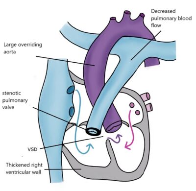
This defect is the most common of the so-called “blue baby” heart defects. It is called Tetralogy of Fallot (TOF) because the French doctor Étienne-Louis Arthur Fallot described it as comprising four abnormalities: 1) The base of the pulmonary artery is narrow. 2) There is a hole in the ventricular septum. 3) The aorta originates more from the right, overriding the septal defect, which means both ventricles pump blood into the aorta. 4) The right ventricle has a thicker-than-normal wall. The thickened wall of the right ventricle is not a separate defect but is due to the elevated blood pressure in the right ventricle.
Because the blood flow to the lungs is impeded, some oxygen-poor venous blood flows from the right ventricle into the aorta, reducing the oxygen saturation of the arterial blood. Some children experience cyanotic spells caused by spasmodic constriction of the pulmonary artery base.
Fallot’s defect is surgically corrected before the age of one year. Pulmonary stenosis is repaired, and the ventricular septal defect (VSD) is closed so that the aorta is entirely on the left ventricular side. The wall thickness of the right ventricle returns to normal as the ventricular pressure decreases. If the pulmonary arteries have become too narrow due to the defect, a shunt surgery is performed first. This auxiliary procedure involves directing additional blood from the aorta at the base of the subclavian artery to the pulmonary artery using a Gore-Tex tube. The increased blood flow enlarges the branches of the pulmonary artery. As pulmonary circulation increases, the oxygen saturation of the arterial blood rises, reducing the cyanosis visible on the tongue and skin. Full correction of the defect is possible once the branches of the pulmonary artery have grown sufficiently large. After corrective surgery, the prognosis for the patient is good, but lifelong cardiac monitoring is required.