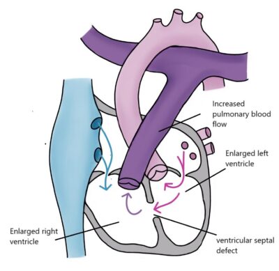
Ventricular Septal Defect (VSD) is an opening in the wall between the heart’s ventricles. It is the most common congenital heart defect. Blood flows through the opening from the left ventricle to the right ventricle. This causes increased blood flow to the lungs and makes the heart’s left ventricle work harder.
Small VSDs, less than three millimeters in diameter, usually do not cause symptoms or problems for a full-term newborn. Many of these close on their own during the first few years of life, so monitoring is often sufficient for management. The size and location of the defect affect the likelihood of closure. Defects located in the muscular (muscular) part of the lower part of the heart’s partition often close on their own.
A large defect can cause symptoms of heart failure in infants as early as a few weeks old. Typical symptoms of heart failure include increased breathing rate and fatigue while feeding. The baby may not feed well and tire quickly during feeding. The child’s liver may also be enlarged, heart rate accelerated, blood pressure and pulse pressure decreased, and growth may be poor. Heart failure caused by shunting is treated with medications, and if necessary, VSD is surgically closed.
Surgical closure of the ventricular septal defect is performed if it continues to affect heart function or the child’s well-being despite medication. If the VSD is large, the left atrium and both ventricles expand, raising the pressure in the right ventricle and pulmonary artery. Elevated pulmonary artery pressure can gradually damage lung vessels and the heart. Timely repair prevents these injuries. Repair surgery is often performed in infancy. Following surgery and recovery, patients lead normal lives.