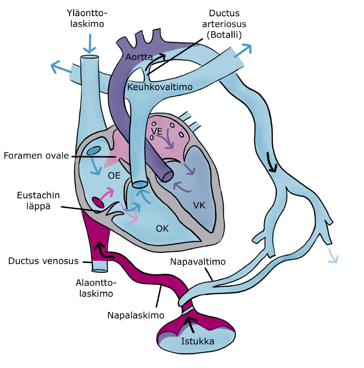
Fetal Circulation
During pregnancy, oxygen and nutrients are transferred to the fetal circulation from the mother’s blood through the placenta. Oxygen-rich, bright red blood flows from the placenta through the umbilical vein to the fetal inferior vena cava. The blood continues to the right atrium, where the Eustachian valve directs the oxygen-rich blood through the oval window (foramen ovale) in the atrial septum to the left atrium. On the left side of this opening in the atrial septum is a valve, which remains open in the fetus. The left ventricle pumps oxygenated blood into the aorta, from which it flows towards various parts of the body, including the brain, which develops rapidly and requires a large amount of oxygen during fetal development.
Deoxygenated, dark red blood returning from the upper body flows through the superior vena cava and the right atrium into the right ventricle, which pumps the blood through the pulmonary artery towards the lungs. Although the fetal lungs are not yet functional, they require a small amount of blood for development. Before the blood reaches the lungs, most of it flows through the open ductus arteriosus into the descending aorta. The deoxygenated blood then continues through the aorta towards the lower body and flows back to the placenta through the umbilical arteries.
Many children with congenital heart defects, where the circulation from the heart to the lungs is inadequate, can appear relatively healthy after birth. This is due to the critical role the placenta plays in fetal circulation. Once the child is born, the umbilical cord is cut, ending the oxygen supply from the placenta. At this point, the child’s condition can deteriorate rapidly.
During the newborn’s first breath, a significant amount of blood flows into the lungs, initiating lung function. In the lungs, the blood binds oxygen from the air, and the increased blood flow to the lungs causes a large volume of blood to flow into the left atrium. The increased blood flow pushes the valve at the opening of the atrial septum (foramen ovale) shut, permanently closing the opening. As the circulation changes, the Eustachian valve atrophies. Within the following days, the ductus arteriosus also closes.
Once the lungs begin to function, the oxygen levels in a healthy newborn’s blood increase. The blood pressure in the aorta rises, while simultaneously, the pressure in the pulmonary blood vessels and the right side of the heart decreases. The decrease in pulmonary arterial pressure continues for several weeks, marking the completion of the child’s circulatory system